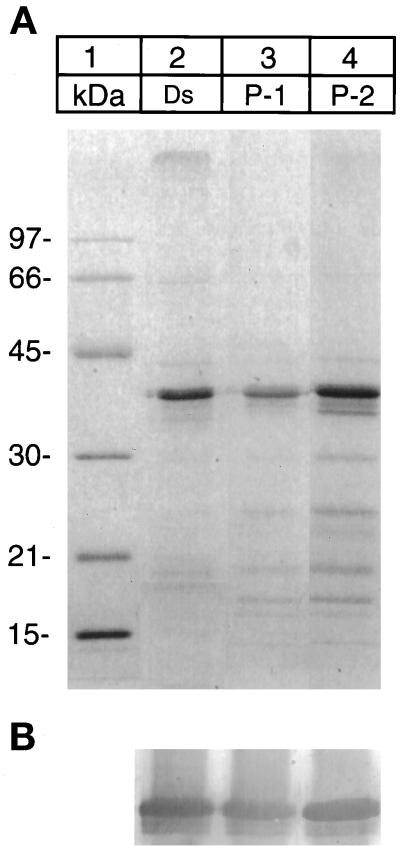
Figure 3.
SDS-PAGE of desaturase preparations isolated from microsomes, P-1, and P-2 fractions. (A) Coomassie Blue-stained gel: lane 2, desaturase (Ds) isolated from microsomes; lane 3, desaturase isolated from P-1 fraction; and lane 4, from P-2 fraction. (B) Immunoblot of the above gel with the antidesaturase antibody as in Figure 1B.

