
Figure 6.
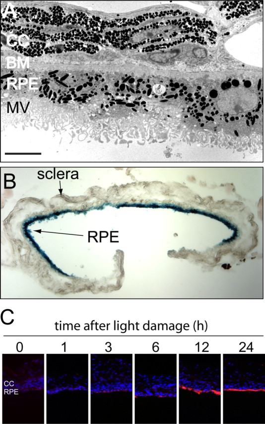
The RPE damage response is maintained in vitro in an eyecup explant. A, Transmission electron microscopy of the RPE and adjacent choroid from a C57BL/6 mouse after 6 d in vitro reveals relatively normal RPE morphology including apical microvilli. CC, Choriocapillaris; BM, Bruch's membrane; MV, microvilli. Size bar, 5 μm. B, RPE cells in an eyecup explant maintain transcriptional and translational competence. An eyecup from a BALB/c mouse was infected in vitro with a recombinant adenovirus vector carrying a β-galactosidase coding region under the control of a cytomegalovirus enhancer/promoter, incubated for 2 d, and then fixed and stained with X-gal. The blue X-gal reaction product is localized to the RPE. C, OSMR expression by RPE cells in eyecup explants prepared at different times after 1.5 h of light damage in vivo. Dark-adapted BALB/c mice were exposed to toxic light, RPE explants were prepared at the indicated times, and after 1 d in vitro the eyecups were immunostained for OSMR.