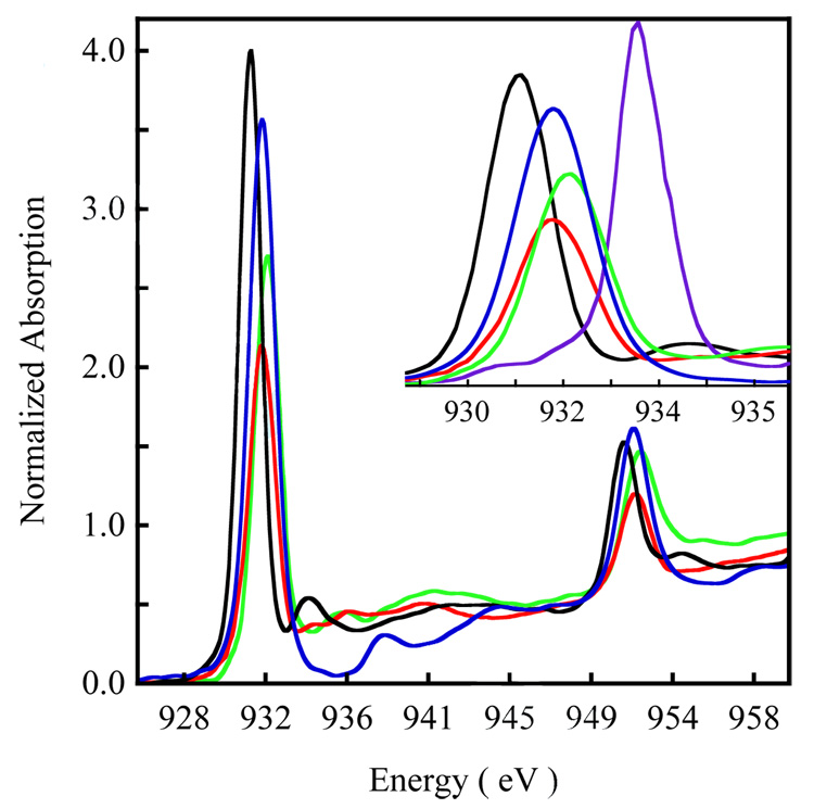
Figure 2.
The normalized Cu L-edge XAS spectrum of 1 ( ), 2 (
 ) and 3 (
 ). The spectrum of D4h [Cu(Cl)4]2− (
 ) has been included as an energy and intensity reference. The inset shows the expanded L3 region. The spectrum of [CuIII(MNT)](TBA) (MNT = maleonitriledithiolate, TBA = tetra-n-butylammonium) 43 has been included for comparison of the ligand-field induced shift in the pre-edge position of a Cu(III) complex.

