FIGURE 3.
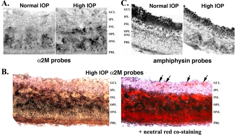
α2-Macroglobulin is preferentially expressed in glia. Retinas were dissected from normal or day 21 glaucoma eyes, and sections were prepared for in situ mRNA hybridization with DIG-labeled probes specific for α2M or Amphiphysin-1. A, normal versus glaucoma retinas labeled with α2M probes. Note the increase in α2M labeling intensity in the glaucoma retina. B, glaucoma retinas labeled with α2M probes, followed by staining with neutral red to show cell bodies (e.g. arrows in the retinal ganglion cell layer, GCL). C, normal versus glaucoma retinas labeled with Amphiphysin-1 probes. Note the reduction in Amphiphysin-1 labeling intensity in the glaucoma retina. GCL, retinal ganglion cell layer; IPL, inner plexiform layer; INL, inner nuclear layer; OPL, outer plexiform layer; ONL, outer nuclear layer; PRL, photoreceptor layer.

