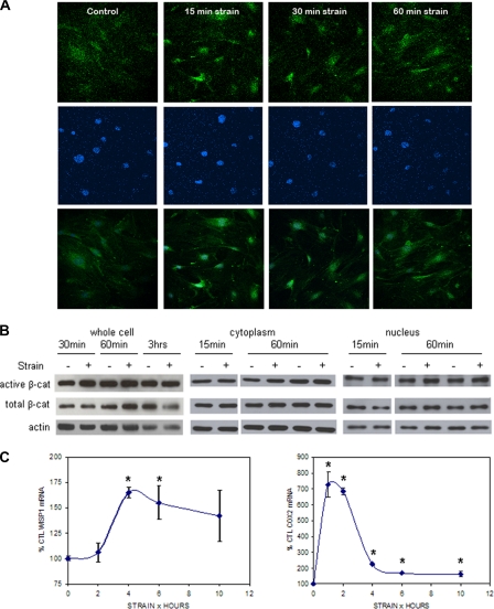
FIGURE 1.
Mechanical strain induces β-catenin accumulation. A, CIMC-4 cells were subjected to strain for 15–60 min and immunostained for active β-catenin (top) and DAPI (middle). Images of active β-catenin and DAPI staining were merged (bottom) to visualize active β-catenin in the nucleus. Increased nuclear active β-catenin was detected 15 min after beginning mechanical strain by confocal microscopy. B, CIMC-4 cells were strained for 15 min to 3 h. Protein from whole cell lysates and cytoplasmic and nuclear fractionates was extracted and run at 10 μg/lane on 7.5% SDS-PAGE. Western blotting showed increases in active and total β-catenin from whole cell lysates at 30 and 60 min, respectively, whereas an increase in both cytoplasmic and nuclear active β-catenin was detected 60 min after strain. Expression of actin is shown as a control for equal protein loading. C, expression of the β-catenin target genes WISP1 and COX2 in CIMC-4 cells 1–10 h after strain initiation was evaluated by real time RT-PCR. A significant increase in WISP1 expression was measured at 4 and 6 h, whereas COX2 expression was increased at all times. Two experiments grouped (mean ± S.E.) for statistical analyses are shown in the graph. *, significant effect of strain (p < 0.05). CTL, control.

