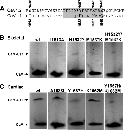
FIGURE 5.
Residues important for CT1-CaM interaction. A, alignment of cardiac and skeletal CT1 in the region analyzed by mutagenesis, with gray shading indicating residues that contact the CaM C-lobe in the crystal structure (21). B and C, non-denaturing PAGE analysis for complex formation between CaM and wild-type (wt) or mutant skeletal or cardiac CT1 in the presence of 2 mm Ca2+.

