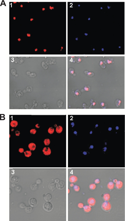
FIGURE 4.
The NES of PKI effectively excludes Hat1p from the nucleus. Subcellular localization of Hat1p was determined by indirect immunofluorescence and confocal microscopy. Cells from strain EMY31 (HAT1-Myc; A) or EMY35 (HAT1-Myc-NES; B) were probed with anti-Myc primary antibody. Hat1p was visualized with a Cy3-conjugated secondary antibody. The images shown are Cy3 (1), 4′,6-diamidino-2-phenylindole (2), differential interference contrast (3), and merge (4).

