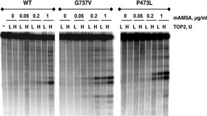
FIGURE 8.
Double strand DNA cleavage by mAMSA-hypersensitive Top2 proteins. Cleavage of linear end-labeled pUC18 DNA was carried out as in the experiment shown in Fig. 7. Samples were treated with proteinase K followed by electrophoresis using 1.5% agarose gels. After electrophoresis, gels were dried and exposed for autoradiography. mAMSA concentrations are indicated in the figure. For each drug concentration, samples with two different amounts of added Top2 protein are shown. Samples marked “L” had 10 units of Top2 added, samples marked “H” had 25 units of Top2 added. For wild-type Top2 and Top2(G737V), 1 unit is 20 ng of protein; for Top2(P473L) 1 unit is 140 ng of protein.

