FIGURE 5.
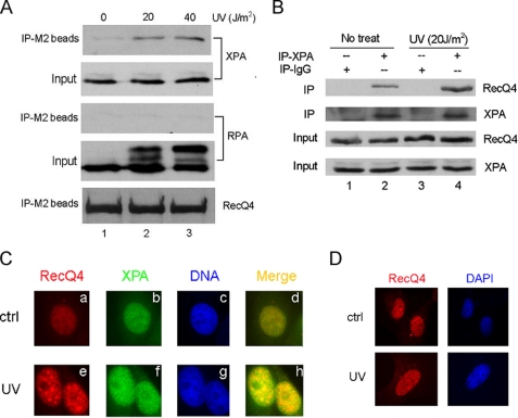
UV irradiation stimulated the interaction between RecQ4 and XPA. A, H1299 cells stably transfected with FLAG-RecQ4 were irradiated with 20 or 40 J/m2 UV or left untreated. At 4 h after UV treatment, whole cell lysates were prepared and immunoprecipitated with anti-FLAG resin (IP-M2 beads) followed by Western blotting with anti-XPA, anti-RPA, and anti-RecQ4 antibodies. 10% of whole cell lysates were used as input. B, HeLa cells were treated with 20 J/m2 UV or left untreated. 4 h after treatment, whole cell lysates were immunoprecipitated with anti-XPA antibody, and precipitated proteins were detected with anti-RecQ4 antibody. 10% of whole cell lysates were also included as input. C, HeLa cells cultured on coverslips were either untreated (upper panels) or treated with 20 J/m2 UV (lower panels). 4 h after treatment, cells were immunostained by anti-RecQ4 (red) and anti-XPA (green) antibodies. The colocalization of RecQ4 and XPA foci were shown in yellow. Nuclei were stained with 4′,6-diamidino-2-phenylindole (blue). ctrl, control. D, XPA-deficient cells cultured on coverslips were either untreated (upper panels) or treated with 20 J/m2 UV (lower panels). 4 h after treatment, cells were immunostained by anti-RecQ4 (red) antibodies. DAPI, 4′,6-diamidino-2-phenylindole.

