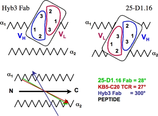
FIGURE 5.
Schematic representation of the positioning of 25-D1.16 and Hyb3 antibodies. 25-D1.16 and KB5-C20 TCR (11) utilize a very similar mode of recognition of MHC-bound peptide that is distinct from that used by Hyb3 antibodies (4). Red and green vectors show virtually identical orientations of 25-D1.16 and KB5-C20 TCR; the dark blue vector indicates a profoundly different orientation of Hyb3; and the black vector indicates peptide positioning in the binding groove of both H2-Kb and HLA-A1 MHC-I proteins.

