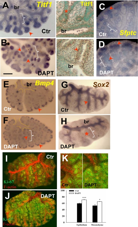
FIGURE 4.
Disruption ofγ-secretase activity expands distal epithelial cell fate and increases distal proliferation during branching. WMISH of Titf1 (A), Sfptc (C), and Bmp4 (E) in control lungs at 48 h shows increased or selective expression of these genes in the epithelium of distal buds (arrowhead) and little to no expression in proximal epithelium (white brackets). These distal marker genes are ectopically expressed in proximal regions of DAPT-treated (50 μm) lungs (arrowhead near white bracket in B, D, and F). Immunostaining of Titf1 (A and B, right) confirms strong ectopic expression in the proximal region of DAPT-treated lungs. G and H, WMISH of Sox2 shows typical expression in proximal epithelium of controls and marked reduction in the Sox2 domain by DAPT (note preserved expression in main bronchi (br)). I and K, immunohistochemistry analysis of Ki-67 (green) and F-actin (red) in controls shows Ki-67 labeling associated mostly with budding regions (terminal and lateral buds; circled area). In DAPT-treated lungs, Ki-67 was increased where ectopic buds formed (compare the F-actin and Ki-67 expression in proximal regions in I and J). J, the increased diffuse Ki67 labeling in the most superficial layer of the proximal mesenchyme partially obscures F-actin signals. K, quantitative analysis of Ki-67-positive cells in budding regions of control and DAPT groups (upper panels, representative field) shows increased labeling in both epithelium and mesenchyme of DAPT (graph shows the percentage of positive cells among total cells; mean ± S.E.; n = 4; *, p < 0.05; **, p < 0.01); all plates are representative of n > 3 explants. Bar (B), 400 μm.

