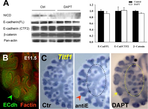
FIGURE 8.
The phenotype of DAPT-treated lungs is not associated with inhibition ofγ-secretase cleavage of E-cadherin. A, Western blot analysis of NICD, E-cadherin (C36 antibody), β-catenin, and panactin in 48 h cultured lungs in which Notch cleavage was blocked by DAPT (50 μm). There is no difference in the pattern of staining or intensity of the full-length E-cadherin (E-Cad/FL), E-cadherin fragment potentially cleaved by γ-secretase (E-Cad/CTF2), or β-catenin. The graph represents densitometry measurements of Western blots (mean ± S.E.; values normalized by panactin; n = 3/group). B, whole mount immunostaining of E-cadherin (green) and F-actin (red) in E12 lung shows strongest E-cadherin signals in distal buds. C, WMISH of Titf1 in lungs cultured in control, E-cadherin blocking antibody (antiE; 100 μg/ml), and DAPT (50 μm)-containing media (48 h). AntiE inhibits branching (arrowhead) but does not result in the enlargement of peripheral buds (yellow arrow) or ectopic budding (circled area) characteristic of DAPT-treated lungs. n = 3/condition.

