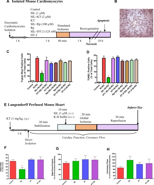
FIGURE 1.
Experimental protocol. A, isolated mouse cardiomyocytes incubated for 1 h with or without sildenafil (SIL) (1 μm) in the presence or absence of protein kinase G (PKG) inhibitor, KT2358 (2 μm), Rp-8-pCPT-cGMPs (100 μm), or DT-2 (125 nm) and subjected to 40 min of SI and 1 or 18 h of RO. Cell necrosis and apoptosis were assessed after 1 or 18 h of reoxygenation, respectively. B, representative image of isolated mouse cardiomyocytes. C, quantitative data showing the effect of sildenafil (1 μm) with or without KT5823 (KT), Rp-8-pCPT-cGMPs (Rp), or DT-2 on cardiomyocyte necrosis following SI-RO as determined by trypan blue staining (* indicates p < 0.001 versus SI-RO; n = 4). D, quantitative data showing the effect of sildenafil (1 μm) with/without KT5823 (KT), Rp-8-pCPT-cGMPs (Rp), or DT-2 on cardiomyocyte apoptosis following SI-RO determined by TUNEL assay (* indicates p < 0.001 versus SI-RO; n = 4). E, isolated perfused hearts subjected to 10 min of intracoronary infusion of sildenafil (1 μm, at 0.25 ml/min pump speed) or Krebs-Henseleit (K-H) buffer followed by 20 min of no-flow global ischemia and 30 min of reperfusion. Myocardial infarct size was determined after the ischemia-reperfusion protocol. KT5823 (1 mg/kg) or volume-matched DMSO (solvent of KT5823) were administered (intraperitoneal) 1 h prior to the heart isolation. F, infarct size (% of risk area); G, double product of heart rate and ventricular developed force (% of pre-ischemic baseline); and H, coronary flow (% of pre-ischemic value). * indicates p < 0.05 versus all other groups.

