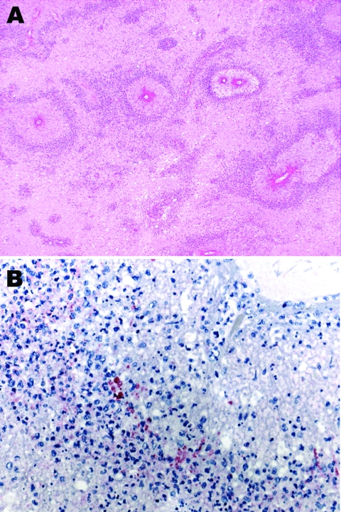
Figure.
A) Subcortical cerebral white matter with numerous perivascular foci of demyelination and necrosis (hematoxylin and eosin stain, original magnification ×40). B) Immunohistochemical evidence of Mycoplasma pneumoniae antigen inside macrophages present in the perivascular inflammatory infiltrate (immunohistochemical assay performed by using the monoclonal anti–M. pneumoniae antibody and naphthol fast red as counterstain, original magnification ×100).

