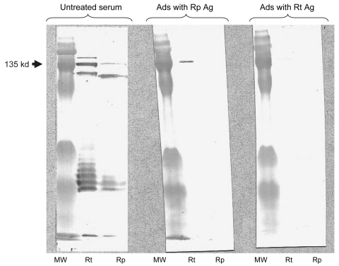
Figure.
Western blot assay and cross-adsorption studies of an immunofluoresence assay–positive serum sample from a patient with rickettsiosis in Algeria. Antibodies were detected at the highest titer (immunoglobulin [Ig] G 256, IgM 256) for both Rickettsia typhi and R. prowazekii antigens. Columns Rp and Rt, Western blots using R. prowazekii and R. typhi antigens, respectively. MW, molecular weight, indicated on the left. When adsorption is performed with R. typhi antigens (column Ads with Rt Ag), it results in the disappearance of the signal from homologous and heterologous antibodies, but when it is performed with R. prowazekii antigens (column Ads with Rp Ag), only homologous antibody signals disappear, indicating that the antibodies are specific for R. typhi.

