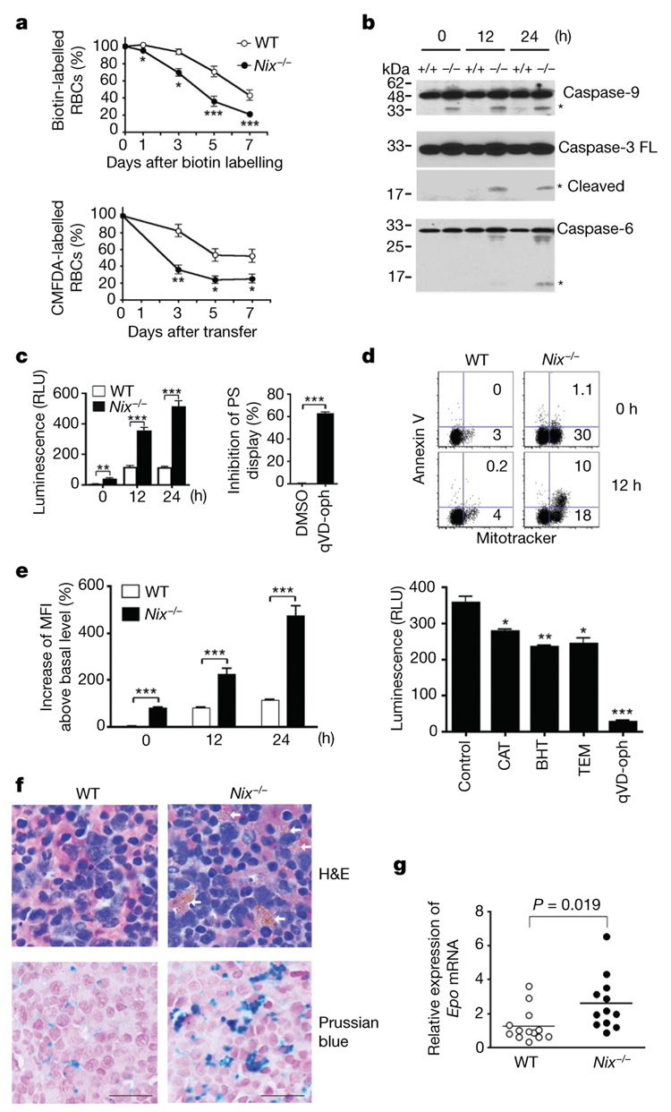
Figure 2. Decreased survival of RBCs in Nix−/− mice.
a, Quantification of NHS-biotin-labelled RBCs or transferred CMFDA-labelled RBCs (n = 3). b, Western blot analyses of caspases in RBCs after in vitro culture. Asterisks denote processed caspases. FL, full length. c, The relative luminescence units (RLU) of caspase activities in cultured RBCs and the suppression of annexin V staining in Nix−/− RBCs after 12-h culture in the presence of qVD-oph or solvent control (DMSO) (n = 3). d, Mitotracker versus annexin V staining of RBCs after in vitro culture. e, Mean fluorescent intensity (MFI) of ROS staining in RBCs after in vitro culture, and caspase activities in Nix−/− RBCs after 24 h culture with solvent control, ROS scavengers or qVD-oph (n = 3). BHT, butylated hydroxytoluene; CAT, catalase; TEM, tempol.
f, Haematoxylin and eosin (H&E) staining of spleen sections of 9-week-old wild-type and Nix−/− mice. Arrows denote iron deposits within macrophage cytoplasm. Iron deposits were also stained with Prussian blue, followed by counterstain with nuclear-fast red. Scale bar, 20 µm. g, Real-time RT–PCR of erythropoietin (Epo) (n = 12). For all relevant panels, statistical significance of the data (mean ± s.e.m.) is: *P<0.05; **P<0.01; ***P<0.001.

