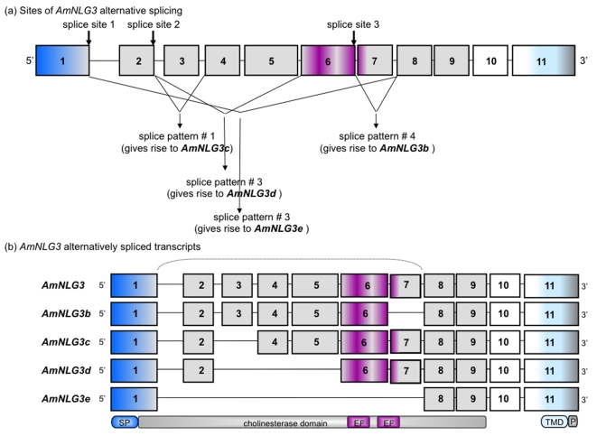
Figure 3. Honeybee Neuroligin 3 Gene Arrangement and Intron/Exon Conservation.
AmNLG3 alternatively spliced transcripts were identified by RT-PCR from honeybee brain cDNA. Arrangement and intron/exon splice sites of AmNLG3 splice variants were deciphered by both NCBI and Beebase BLAST tools against genomic DNA. Donor/acceptor splice sites were confirmed using NCBI SPIDEY. Sizes of exons, as well as intron gaps, are not drawn to scale. Exons are numbered from 5′ to 3′. (a) highlights the three sites of alternative splicing and resulting splice patterns. (b) illustrates the five alternatively spliced AmNLG3 transcripts which correspond to the splicing patterns show in (a). The bracket highlights that splicing occurs within the cholinesterase domain. Signal peptide, cholinesterase, transmembrane domains and EF hand metal and PDZ binding motif are drawn below the encoding exons. Abbreviations SP: signal peptide; EF: EF hand metal binding motif; TMD: trans-membrane domain, P: PDZ binding domain.

