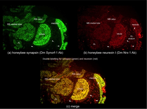
Figure 8. Honeybee Neurexin I Protein Expression in the Brain.
Immuno-staining of forager brain sections (a) for synapsin using SynOrf-1 antibody and incubation with Alexa-488-conjugated anti-mouse antibody allowing green colouration to highlight protein expression; (b) for honeybee neurexin-I using DmNrx-1 antibody and incubation with Alexa-546-conjugated anti-mouse antibody allowing red colouration to highlight protein expression; (c) merge shows neurexin-I and synapsin co-localise in mushroom body neuropil (MB medial lobe and MB calyx), optic lobe neuropil (medulla and lobulla) and antennal lobes. Small puncta of neurexin I expression, distinct to synapsin, are highlighted by small arrow heads within the optic lobe (OL) stratum.

