Fig. 3.
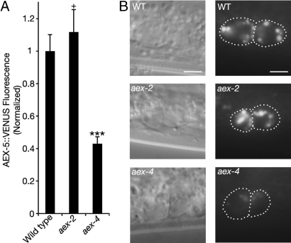
Intestinal aex-4 regulates the secretion of AEX-5. (A) In aex-4 mutants, the total fluorescence, normalized to WT, of intestinal AEX-5::VENUS in coelomocytes is significantly less than that of WT. There is no significant change in coelomocyte fluorescence in aex-2 mutants. ***, P < 0.0005 significantly different from the respective mutant. +, P > 0.05 not significantly different from WT. Error bars represent SEM. (B) Representative photographs of anterior coelomocytes in WT, aex-2, and aex-4. The left image is a Nomarski image. The right image is AEX-5::VENUS fluorescence. (Scale bars, 5 μm.)

