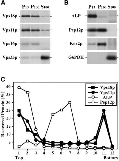
Figure 8.
Subcellular localization of the class C Vps proteins. (A) Wild-type spheroplasts (SEY6210) were labeled with Expres35S for 30 min and chased for 60 min before lysis in a hypotonic buffer. After centrifugation at 300 × g, the lysate was subjected to sequential centrifugation to generate 13,000 × g pellet (P13), 100,000 × g pellet (P100), and 100,000 × g supernatant (S100) fractions. The presence of Vps18p, Vps11p, Vps16p, and Vps33p in each fraction was determined by quantitative immunoprecipitation, SDS-PAGE, and fluorography. (B) The distribution of several organelle marker proteins in these fractions was also determined by immunoprecipitation: mALP (vacuole), Pep12p (endosomal compartment), Kex2p (late Golgi), and glucose 6-phosphate dehydrogenase (G6PDH, cytoplasm). (C) Accudenz gradient fractionation of cell lysates. Wild-type spheroplasts (SEY6210) were labeled for 30 min with Expres35S and chased for 60 min before lysis in a hypotonic buffer. The lysate was cleared at 300 × g and was then loaded on top of an Accudenz gradient. After centrifugation to equilibrium, gradient fractions were collected and Vps18p, Vps11p, ALP, and Pep12p were recovered by immunoprecipitation and resolved by SDS-PAGE.

