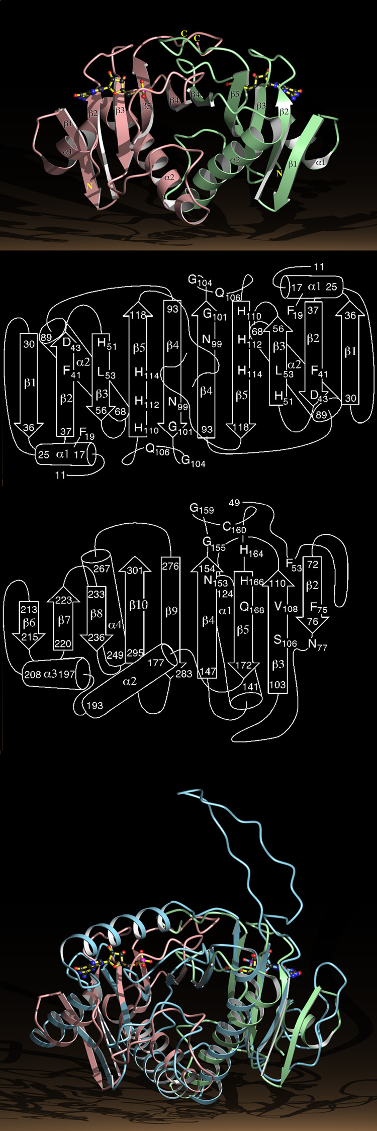
Fig. 3.
Structure of the HINT Dimer and the GalT Monomer.
a, Ribbon diagram of the HINT-GMP dimer with numbered secondary structural elements. The right hand HINT monomer is in green, the left in pink.
b, Secondary structure of the HINT dimer with the positions of a subset of the conserved residues in the HIT superfamily.
c, Secondary structure of residues 49 to 302 of the GalT core identifying residues that align in three dimensions with HINT.
d, The Cα positions of residues 49 to 302 of the GalT core, represented by a ribbon diagram in blue, superimposed on the HINT-GMP dimer.

