Figure 5.

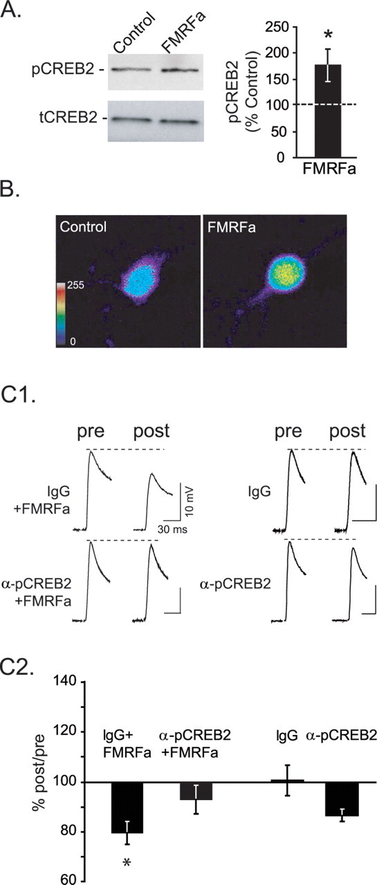
CREB2 is phosphorylated after FMRFa treatment and is necessary for FMRFa-induced LTD. A, Immunoblot analysis using the anti-phospho-CREB2 antibody showed that immediately after the end of treatment with FMRFa, levels of pCREB2 were significantly increased compared with control. Exposure to FMRFa did not affect levels of total CREB2 (*p < 0.05). B, Under control conditions, pCREB2 was observed primarily in the nucleus of cultured isolated sensory neurons and to a lesser extend in the cytoplasm, displaying a distribution pattern that is similar to that of mammalian CREB2 in rat cultured cortical neurons (White et al., 2000). After FMRFa exposure, an increase in pCREB2 was observed in both the cytoplasm and nucleus, suggesting that the presence of a postsynaptic target is not required for CREB2 to be phosphorylated. C1, Representative EPSPs recorded from IgG-injected (top traces) or phosphospecific anti-CREB2-injected (bottom traces) cocultures, before and after treatment with FMRFa or vehicle. C2, For statistical analysis, the peak amplitude of EPSP at the 24 h posttest was normalized to pretest EPSP. Injection of anti-phospho-CREB2 antibody blocked LTD (*p < 0.05) without significantly affecting basal transmission. Error bars indicate SEM.
