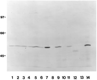
Figure 1.
Expression of fascin by cell lines used in attachment assays. Equivalent numbers of cells were lysed in SDS-PAGE sample buffer, the extracts were resolved on a 12.5% polyacrylamide gel under reducing conditions, transferred to nitrocellulose, and probed with antibody 55K2 to fascin. Lanes: 1, A10 cells; 2, HISM cells; 3, MG-63 cells; 4, SK-N-SH cells; 5, H9c2 cells; 6, G8 cells; 7 and 14, C2C12 cells; 8, A549 cells; 9, C32 cells; 10, RT4 cells; 11, HT1080 cells; 12, G361 cells; 13, MDCK cells. Molecular mass markers are indicated in kDa.

