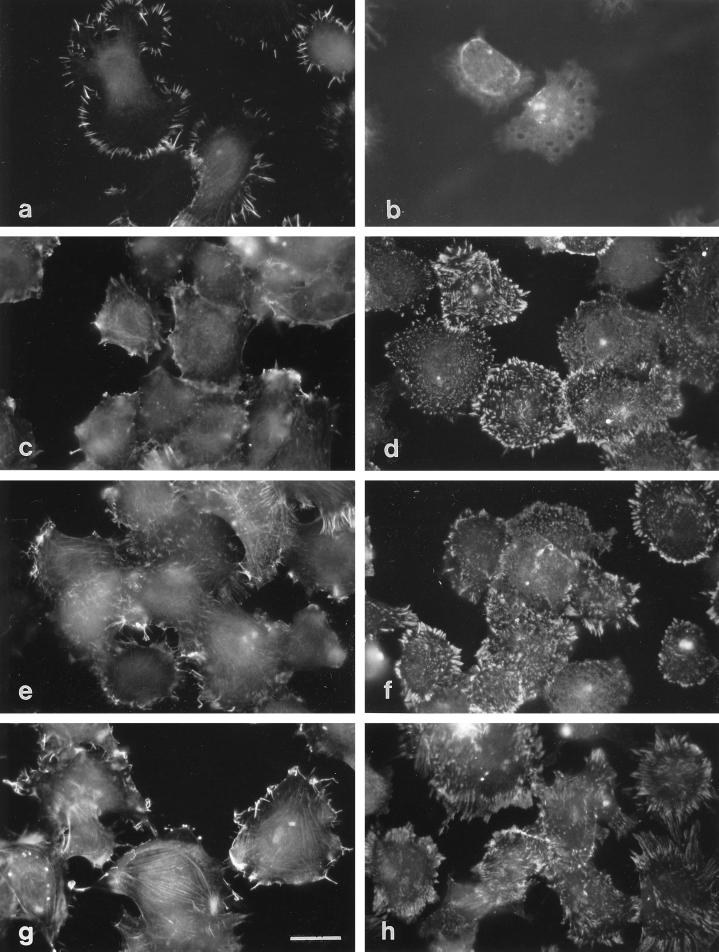
Figure 3.
Formation of fascin microspikes and focal contacts by H9c2 cells plated on various ECM glycoproteins. H9c2 cells were stained for fascin (a, c, e, and g) or vinculin (b, d, f, and h) 1 h after plating on substrata coated with 50 nM TSP-1 (a and b), vitronectin (c and d), collagen IV (e and f), or EHS laminin (g and h). Bar, 12 μm.

