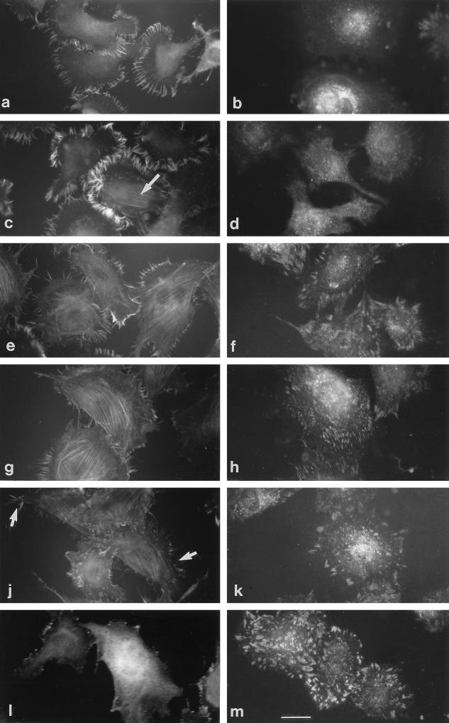
Figure 4.
Formation of fascin microspikes and focal contacts by H9c2 cells adherent on mixed TSP-1/fibronectin substrata. H9c2 cells were stained for fascin (a, c, e, g, j, and l) or vinculin (b, d, f, h, k, and m) 1 h after plating on substrata consisting of 100% TSP-1 (a and b), 90% TSP-1:10% fibronectin (c and d), 75% TSP-1:25% fibronectin (e and f), 50% TSP-1:50% fibronectin (g and h), 25% TSP-1:75% fibronectin (j and k), or 100% fibronectin (l and m). A cell displaying weak colocalization of fascin with microfilaments is shown with an arrow in c. Residual isolated microspikes are shown with arrows in j. Bar, 10 μm.

