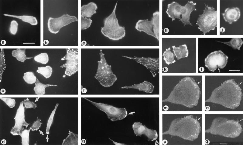
Figure 8.
Presence of fascin-positive microspikes and ruffles at the protrusive margins of migratory and postmitotic C2C12 cells. Sparse cultures of C2C12 myoblasts were stained with rhodamine-phalloidin (a, b, and e), antibody to vinculin (c and f), or antibody to fascin (d, g, and h–q) and examples of small polarized cells (a, b, c, and d), larger fan-shaped cells (e, f, and g), or postmitotic pairs of cells (h–l) photographed under epifluorescence. Arrows in d and g indicate fascin microspikes and ruffles at leading edges; arrowhead in g indicates postmitotic pair of cells. (h) Asymmetric pair of cells nearing completion of cytokinesis which bear fascin microspikes and ruffles at their protrusive margins. (j and l) Examples of cells at the onset of cytokinesis: arrow in l indicates cleavage furrow. (k) Example of postmitotic pair stained 2 h after release from nocadazole block. Bars: a and l, 10um. (m–q) Series of projections from 3D reconstruction of confocal serial optical sections through one cell of a postmitotic pair stained for fascin. Projections were made at 180° (m), 210° (n), 230° (p), and 250° (q) to the original plane of section. Arrows indicate an upraised group of fascin microspikes displayed by the projections. Bar, 2.5 μm.

