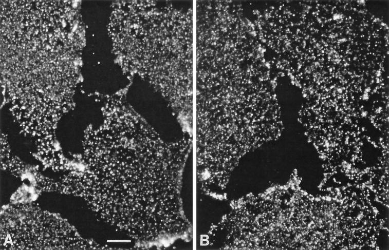
Figure 2.
Immunofluorescence of plasma membranes performed with antibodies specific for G protein subunits. En face views of the inner side of plasma membrane fragments were obtained by sonicating MA104 cells adherent to coverslips. Oregon green-conjugated secondary antibodies were used to visualize primary antibodies to detect: (A) αi subunits with B087 antibodies (10 μg/ml) or (B) β subunits with T20 antibodies (1 μg/ml). The larger areas of the photographs that are devoid of fluorescent signal represent spaces where plasma membrane fragments are absent. Scale bar, 2 μm.

