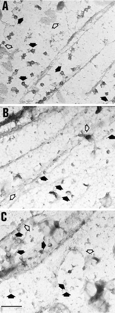
Figure 7.
Immunogold labeling of plasma membranes with caveolin or αi antibodies. Electron micrographs show en face views of the inner side of plasma membrane fragments that have been torn from the upper surface of cultured fibroblasts. The location of some of the morphologically identifiable caveolae (full or partial doughnut shapes indicated by closed arrows) and coated pits (flat or curved honeycomb patterns indicated by open arrows) are shown. (A) Caveolae are decorated well by gold particles when the polyclonal (panel A, 1 μg/ml) or monoclonal (not shown) caveolin antibodies are utilized. (B) Morphologically identifiable caveolae are infrequently labeled by αi reactive B087 antibodies (10 μg/ml) or (C) A569 antibodies (10 μg/ml). The letters at the upper left corner of each panel are placed over a small area that is devoid of plasma membrane or gold particles. Scale bar, 0.5 μm.

