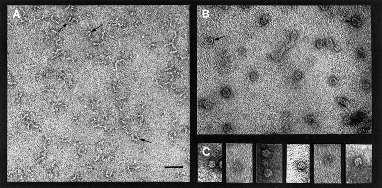
Figure 2.
Electron microscopy of negatively stained RanBP2. Purified RanBP2 was adsorbed to glow-discharged carbon-coated grids and was negatively stained with uranyl acetate. Panels A and C are electron micrographs of two different grid preparations that show predominantly an extended (panel A) or folded (panel B) conformation of RanBP2. Arrows indicate examples of partially folded conformations of RanBP2 found in both preparations. Panel C is a gallery of folded RanBP2 molecules selected from the same preparation as panel B. Bar, 100 nm.

