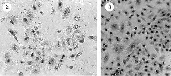
Figure 3.
184A1 growth in TGFβ at different passages. Giemsa-stained cells labeled with [3H]thymidine for 24 h as described in MATERIALS AND METHODS. (a) Small slowly growing area at p28, part of a slightly larger area, labeled after 14 d exposure to TGFβ; this represents the best growth seen at p28. (b) Representative area of a typical p44 colony with good growth (LI >50%) labeled after 13 d exposure to TGFβ. Magnification, 125×.

