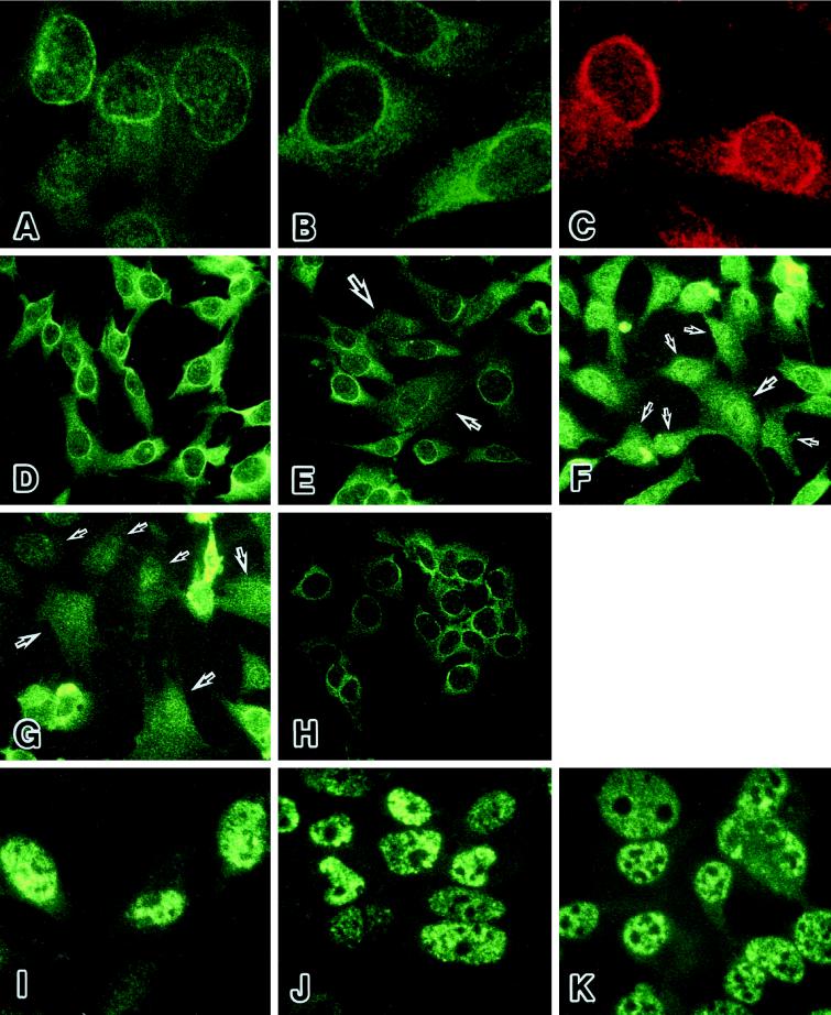
Figure 5.
The localization of Oct-1 protein in nuclear periphery during cellular senescence and immortalization. (A-C) The nuclear periphery of HuS-L12 cells at PDL 66 or 73 was prominently stained by three independent anti-Oct-1 antibodies. (A) YL15, a monoclonal antibody against the unique POU-domain linker region of Oct-1. (B) YL123, a monoclonal antibody against the POU homeodomain of Oct-1. (C) A polyclonal antibody against the C terminus of Oct-1. (D–H) The prominent signals of Oct-1 protein disappeared from the nuclear periphery during cellular aging and were resumed in immortalization. The Oct-1 protein was stained with YL15 in HuS-L12 cells at PDL 66 (D), 73 (E), 84 (F), and 91 (G) and in immortalized IML12–4 cells (H). Arrows indicate the cells that lack the Oct-1–associated nuclear peripheral structure. (I–J) The staining patterns of p53 were mostly unchanged as a control. The p53 protein was stained with pAb421 in HuS-L12 cells at PDL 66 (I) and 91 (J) and in immortalized IML12–4 cells (K).

