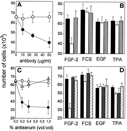
Figure 9.
Effect of anti-αvβ3 antibodies on the mitogenic activity of soluble FGF-2. (A) GM 7373 cells grown on tissue culture plates were incubated with FGF-2 (10 ng/ml) in the presence of the indicated concentrations of monoclonal antiαvβ3 antibody (black symbols) or irrelevant antibody (open symbols). (B) Cells were treated with 10% FCS, EGF (30 ng/ml), or TPA (100 ng/ml) in the absence (black bars) or in the presence of 50 μg/ml of monoclonal anti-αvβ3 antibody (white bars) or of irrelevant antibody (gray bars). (C) Cells grown on tissue culture plates were incubated with FGF-2 (10 ng/ml) in the presence of the indicated concentration of anti-αvβ3 antiserum (•), antiα5β1 antiserum (O), or irrelevant antiserum (Δ). (D) Cells were treated with 10% FCS, EGF, or TPA (doses as in panel B) in the absence (black bars) or in the presence of 1% (vol/vol) of anti-αvβ3 antiserum (white bars), anti-α5β1 antiserum (gray bars), or irrelevant antiserum (striped bars). After 24 h, all cell cultures were trypsinized and cells were counted in a Burker chamber. Control GM 7373 cells incubated with 0.4% FCS or 10% FCS in the absence of any addition were 37,000 ± 7,000 and 72,500 ± 5,600 cells/well, respectively. Each point is the mean ± SEM of two to three determinations in duplicate.

