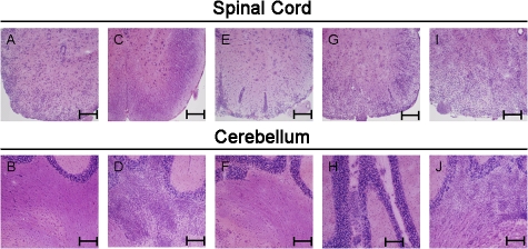
Figure 3.
IFN-γ production in a fraction of pathogenic T cells protects the cerebellum from inflammation. C57BL6/J mice received either 5 × 10⁶ MOG35-55-specific Th1 cells generated in C57BL6/J (WT Th1; A and B), 5 × 10⁶ MOG35-55-specific Th1 cells generated in IFN-γ–deficient (IFN-γ KO) mice (C and D), 5 × 10⁶ WT and 5 × 10⁶ IFN-γ KO (E and F), or 106 WT and 5 × 10⁶ IFN-γ KO (G–J) as a single i.v. injection. 17 d after injection and after development of classical EAE (A, B, and E–H), nonclassical EAE (C and D), or both classical and nonclassical disease (I and J), spinal cord and cerebellum were collected and stained with H&E to reveal inflammation. Representative slides from five independent experiments are shown. Bars, 200 μm.

