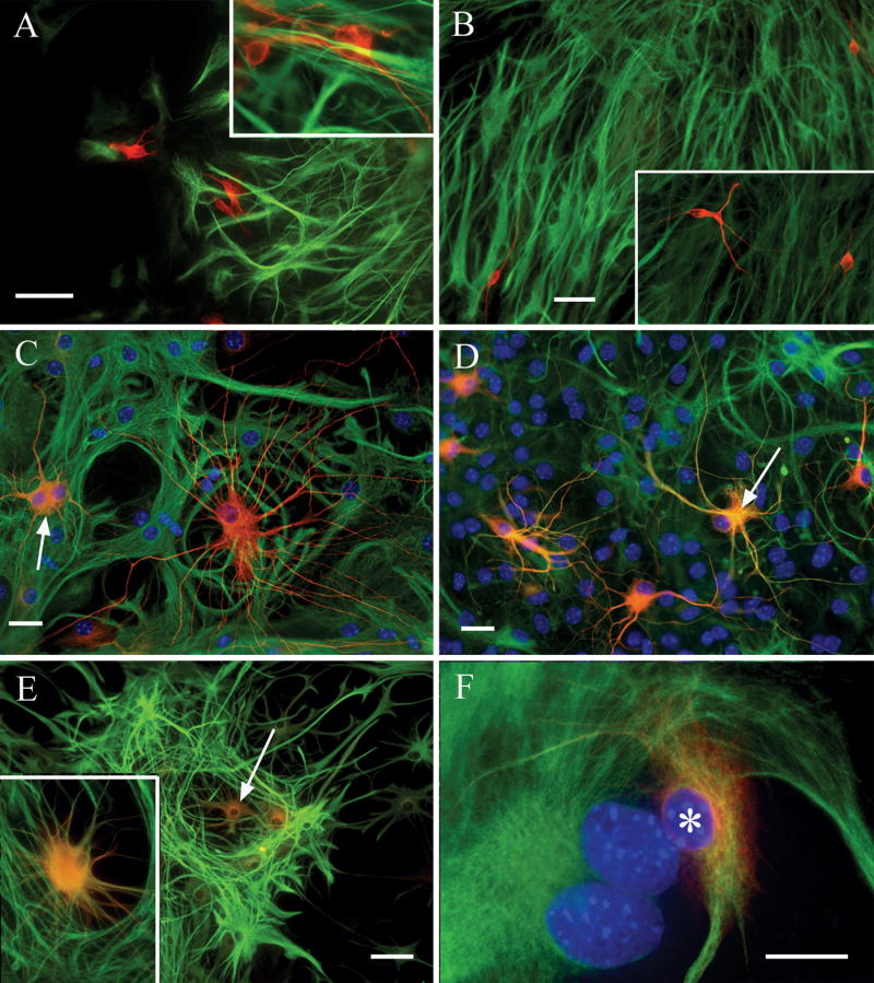
Figure 1. Combined β-III tubulin and GFAP immunolabeling reveals the temporal progression of the asteron phenotype.
Shortly after initiation of differentiation (A), spheres contain non-overlapping populations of cells immunopositive for either β-III tubulin (red), or GFAP (green). No cells co-expressing both markers are seen (inset in A shows higher magnification of non-overlapping neuron/astrocyte labels). Note also the relatively short, poorly branched neuronal processes at this early time point. By twenty-four hours post-plating (B), spheres still contain cells with mutually exclusive β-III tubulin (red)/GFAP (green) labeling patterns, but neuronal morphology is more mature, showing longer processes with greater branching (inset in B). Two days after plating (C), phenotypic heterogeneity is increased, with some cells co-labeling with both neuron (red) and astrocyte (green) markers (arrow points to a co-expressing “yellow” cell). Additionally, cells immunolabeled exclusively with β-III tubulin can be seen displaying “hybrid” morphologies that combine fine, highly branched processes with wide, flat somas (i.e. the large red cell in C). At three days post-plating (D), few cells exclusively express β-III tubulin, and the number of co-expressing asterons increases (e.g. arrow). Asterons at this stage also begin to acquire more “astrocytic” morphologies, with thicker processes and less rounded somas. At five days (E) neurons are scarce and co-expressing asterons (e.g. arrow) display astrocytic morphologies. Inset in (E) shows a higher magnification of an asteron at this stage. By six days (F) co-expressing cells show a wide, flat, fibroblast-like morphology typical of cultured astrocytes (asterisk). Blue in panels C,D,F is Hoechst 33342 nuclear counterstain. Scale bar = 50 uM in (A), 25 uM in (B), and 20 uM in (C&D).

