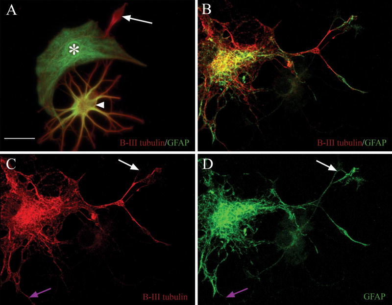
Figure 2. Antibodies against β-III tubulin and GFAP reveal three immunophenotypes, and label separate sub-cellular elements within asterons.
In (A) β-III tubulin+ neurons (red, arrow), GFAP+ astrocytes (green, asterisk), and co-expressing asterons (arrowhead) can be seen in close proximity with no evidence of antibody cross-reactivity in either the exclusively red or green cells. Confocal microscopy (B-D) shows the co-localization of both immunomarkers in a single z-axis of an asteron. Notice that the red and green chromagens often label separate intracellular structures. Some areas labeled with green are unlabeled with red (compare white arrows in C & D), and vice versa (compare purple arrows in C & D). Scale bar = 20 uM in all panels.

