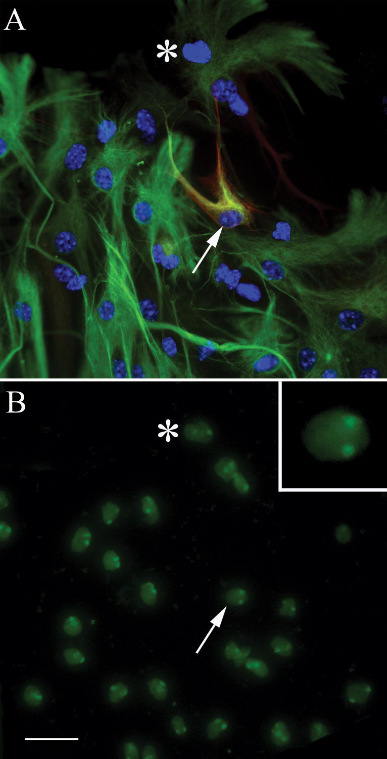
Figure 6. Chromosome painting reveals that asterons are diploid.
Asterons from female mouse cerebellar spheres, defined by co-expression of β-III tubulin and GFAP (red & green, respectively in A), are shown to be diploid when hybridized with probes specific for mouse x-chromosome (green in B). The same general field is shown before (A) and after (B) FISH chromosome painting. Asterisk is added for reference. The asteron indicated by the arrow in (A) can be seen to have two x-chromosomes in (B). Inset in (B) shows a higher magnification of this cell. Scale bar= 25 uM.

