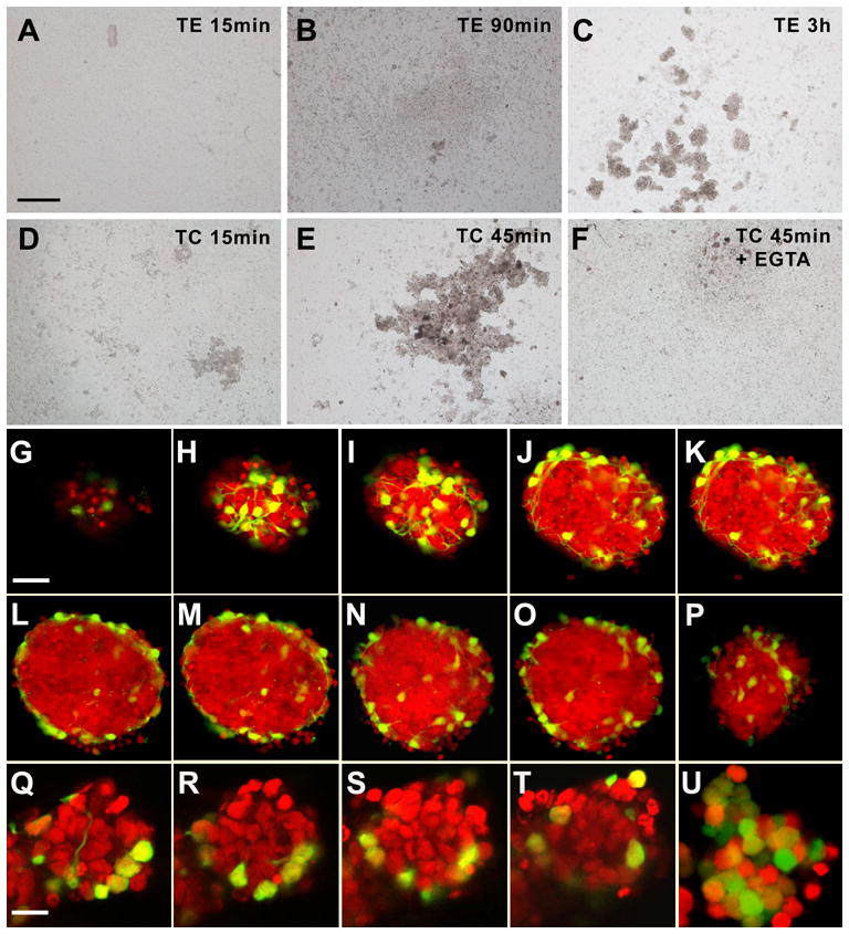
Figure 6. Cell segregation in cerebellar reaggregate cultures prepared from P8 cerebella.

(A–F) Phase contrast images from cultures prepared with cells trypsinized under cadherin-sparing conditions (D–F) or conditions under which all surface cadherins like other surface proteins are digested (A–C).When cadherins are spared, reaggregates may be detected after 15 min or 45 min (D,E). This may be blocked by calcium withdrawal (F; Ca2+ was chelated with EGTA).When cell cadherins were destroyed during trypsination, reaggregation was not seen after 15 min or 90 min, but only after some 3 h. Bar (in A, for A–F) = 200 μm.
(G–P) Optical sections through a characteristic reaggregate culture after 48 h in vitro. After fixation and RNA digestion, nuclei were stained with propidium iodide. Optical sections were sampled every 3 μm, and every other section is shown. Pax2-GFP-positive cells are located preferentially on the surface of the aggregate. Note that G is a top view of the aggregate, and panels H and I are tangential sections close to the upper pole. P is a tangential section close to the basal pole of the aggregate. Bar = 20 μm (in G, for G–J)
(Q–T) Series of optical sections through a 90 min old culture of cells trypsinized under cadherin-sparing conditions. Optical sections were sampled every 2.5 μm, and every other section from the equatorial part of the aggregate is shown. Again, Pax2-GFP-positive cells are preferentially at the margin/surface of the aggregate.
(U) This control aggregate, obtained from a cerebellum of a Math1-EGFP transgenic mouse, documents that cells in the center of aggregates are of the granule cell lineage. Bar = 10 μm (in Q, for Q–U)
