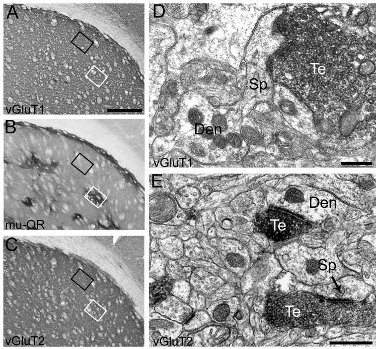
Figure 2. Distribution of vGluT1- and vGluT2-immunolabeled axon terminals in the patch and matrix compartments.
Patch and matrix compartments of the striatum were identified by immunostaining for μ-OR at the light microscopic level (B). Sections immediately preceding and after that stained for μ-OR were incubated with vGluT1 or vGluT2 antibodies and processed for electron microscopic observation. Immunostaining for vGluT1 (A) and vGluT2 (C) at the light microscopic level is shown here to illustrate how blocks of tissue from the patch and matrix were obtained. Sections stained for vGluT1, μ-OR, and vGluT2 were placed side-by-side and areas corresponding to the patch (white box) and matrix (black box) in the μ-OR-immunostained section were cut out form the vGluT1- and vGluT2-immunolabeled sections. At the electron microscopic level, vGluT1- and vGluT2-immunoreactivity was restricted primarily to axon terminals (D-E) forming asymmetric synapses (arrow in E). Scale bar, shown in A, represent 500μm for all light microscopic images. Scale bar in D-E represent 0.5μm Te: terminals; Den: dendrite; Sp: spine.

