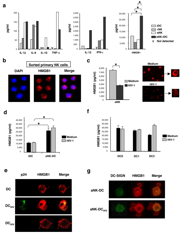
Figure 2. aNK-DC cross-talk triggers HMGB1 expression in both aNK cells and DCs.
(a) 24 h cell-free culture supernatants of iDCs, rNK cells, aNK cells (106/ml), or cocultures of aNK cells and iDCs (ratio 1∶5) were tested for cytokine content. MAP technology was used to quantify IL-1β, IL-6, IL-10, TNF-α, IL-12 and IFN-γ, whereas HMGB1 was quantified by ELISA. * p<0.05 (non-parametric Mann-Whitney test). (b) HMGB1 expression was detected by immunofluorescence (in red) in freshly sorted blood NK cells. Counterstaining with DAPI (in blue) showed the nuclear localisation of HMGB1. (c) Incubation of aNK cells with HIV-1 inhibits HMGB1 secretion. Left panel: aNK cells (106 cells/ml) were incubated in medium or with HIV-1BaL (1 ng/ml of p24) for 3 h and tested for HMGB1 production 21 h later. Data represent three independent experiments and values are means±sd. Right panel: immunofluorescence analysis of HMGB1 expression in the same preparations of aNK cells. (d) HMGB1 production during aNK-iDC cross-talk is not inhibited by HIV-1 infection of iDCs. iDCs were incubated for 3 h in medium or with HIV-1BaL (1 ng/ml of p24) and further cocultured for 21 h with aNK cells (aNK∶iDC ratio 1∶5). HMGB1 concentration was then measured in culture supernatants. Data represent the mean±sd of three independent experiments. (e) Immunofluorescence confocal analysis of HMGB1expression in uninfected or HIV-1-infected iDCs. Upper panel: non infected iDCs; middle panel: HIV-1-infected and replicating iDCs, as shown by intracellular p24 staining; lower panel: iDCs incubated with HIV-1 but negative for intracellular p24 expression. (f) Mature DCs were generated by 48 h stimulation of iDCs with LPS (DC0), soluble CD40L (DC1) or LPS+PGE2 (DC2). DC0, DC1 and DC2 were incubated for 3 h in medium or infected with HIV-1BaL (1 ng/ml of p24) and further incubated in medium for 21 h. HMGB1 quantification in culture supernatants was performed. The mean±sd of three independent experiments is shown. (g) Immunofluorescence analysis of HMGB1 expression in conjugates of aNK cells and uninfected (upper panel) or HIV-1-infected DCs (lower panel) in a 24 h coculture. DCs are DC-SIGN+ and both aNK cells and DCs express HMGB1 in these conjugates. Pictures from one representative experiment out of three conducted with different primary cell preparations are shown.

