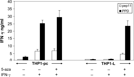
Fig. 6.
HLA-DR2-restricted, antigen-specific T cell activation assay using THP-1 as APC. THP1-L was treated with 5-aza for 4 days; after this period, both THP1-L and THP1-pc cells were treated with IFN-γ for 6 h and then extensively washed to remove traces of the cytokine. Cells were pulsed with antigen (either peptide pep11 or whole PPD protein) and then mixed with a pep11-specific CD4+ T cell clone in flat-bottomed microtiter plate at a 2:1 stimulator/responder ratio. Activation of T cell was quantitatively assessed after 20 h of co-culture by measuring the amount of IFN-γ secreted by ELISA.

