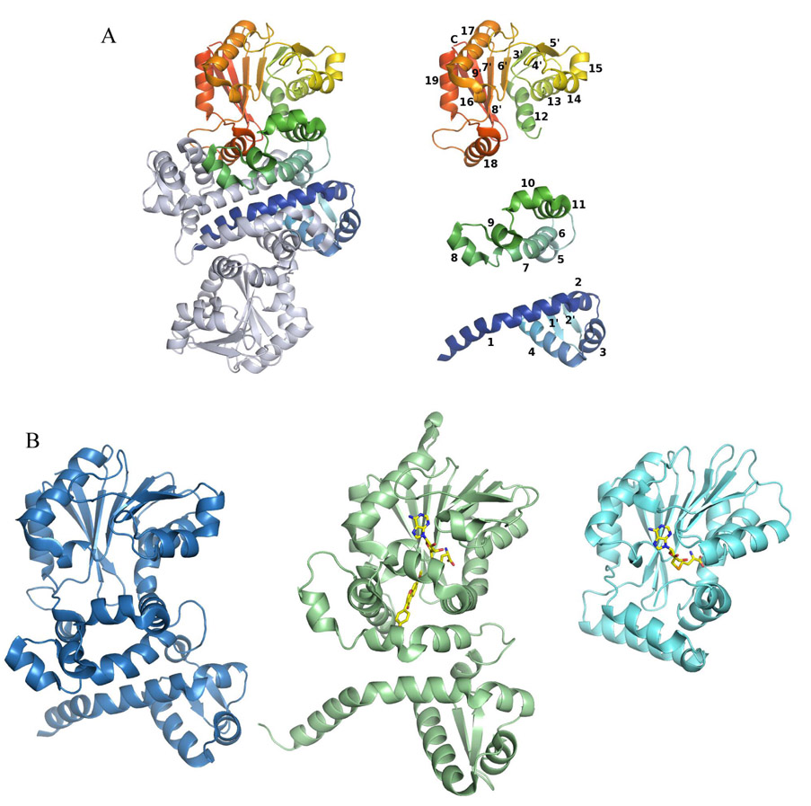
Figure 2.
Structure of the PhzM dimer, illustration of the secondary structural elements of PhzM, and a comparison of the PhzM monomer with representative methyltransferases. (A) The PhzM dimer, one subunit in rainbow coloration, the other in gray. Secondary structure diagram of the PhzM monomer separated by domain and colored as in the dimer. Helices are numbered 1–18, strands are numbered 1'–9'. (B) Ribbon diagrams comparing the structure of PhzM with those of a structural homolog (IOMT), and a typical monomeric small molecule methyltransferase, YecO, from Heamophilus influenzae.

