Figure 2.
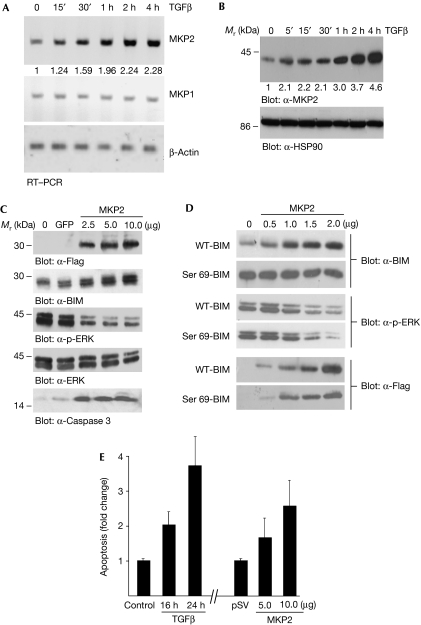
Induction of the MAPK phosphatase MKP2 by TGFβ. (A) AML-12 cells were treated for the indicated times with TGFβ. RNA was isolated and RT–PCR analysis was carried out with primers specific for MKP2, MKP1 and β-Actin; β-Actin acts as a loading control. (B) WCLs were prepared and analysed by immunoblotting; α-HSP90 blot acts as a loading control. RT–PCR gels (A) and immunoblots (B) were quantified as described in the Methods and the fold change over control untreated cells is shown below each corresponding band. (C) Flag-tagged MKP2 (2.5, 5 and 10 μg DNA) was transfected into COS7 cells (GFP was used as a control transfected DNA) and after 24 h incubation WCLs were analysed by immunoblotting for Flag-MKP2, BIM, p-ERK, total ERK and cleaved caspase 3. (D) COS7 cells were co-transfected with 2.0 μg of either WT BIM or the phosphorylation mutant, Ser 69-BIM, and increasing concentrations (0.5, 1, 1.5 and 2 μg) of MKP2. WCLs were prepared and analysed by immunoblotting. (E) AML-12 hepatocytes were either treated with TGFβ for 16 or 24 h, or transfected with MKP2 (5 and 10 μg) for 24 h and apoptosis was quantified by ELISA. The mean±s.d. of three independent experiments each performed in duplicate (n=3) is shown. AML, acute myeloid leukaemia; ELISA, enzyme-linked immunosorbent assay; GFP, green fluorescent protein; HSP90, heat shock protein 90; IL-3, interleukin-3; MAPK, mitogen-activated protein kinase; RT–PCR, reverse transcription–PCR; TGFβ, transforming growth factor-β; WCL, whole cell lysate; WT, wild type.

