Figure 4.
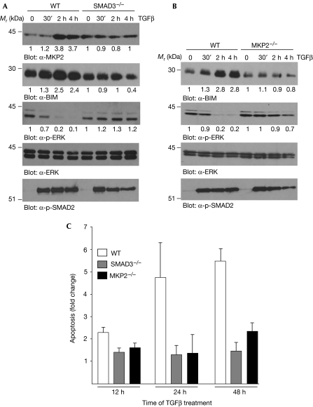
TGFβ-mediated effects in wild-type, SMAD3- and MKP2-deficient pro-B lymphocytes. (A) Bone marrow-derived pro-B cells isolated from WT and SMAD3-deficient (SMAD3−/−) mice were treated with TGFβ for the indicated times, and WCLs were analysed by immunoblotting. (B) Pro-B cells isolated from WT and MKP2-deficient (MKP2−/−) mice were treated with TGFβ for the indicated times, and WCLs were analysed by immunoblotting. Immunoblots of (A,B) were quantified as described in the Methods and the fold change over control untreated cells is shown below each corresponding band. (C) WT and MKP2−/− pro-B cells were treated with TGFβ for 16 and 24 h, and apoptosis was quantified by ELISA. The mean±s.d. of three independent experiments each performed in duplicate (n=3) is shown. ELISA, enzyme-linked immunosorbent assay; TGFβ, transforming growth factor-β; WCL, whole cell lysates; WT, wild type.

