
FIGURE 3.
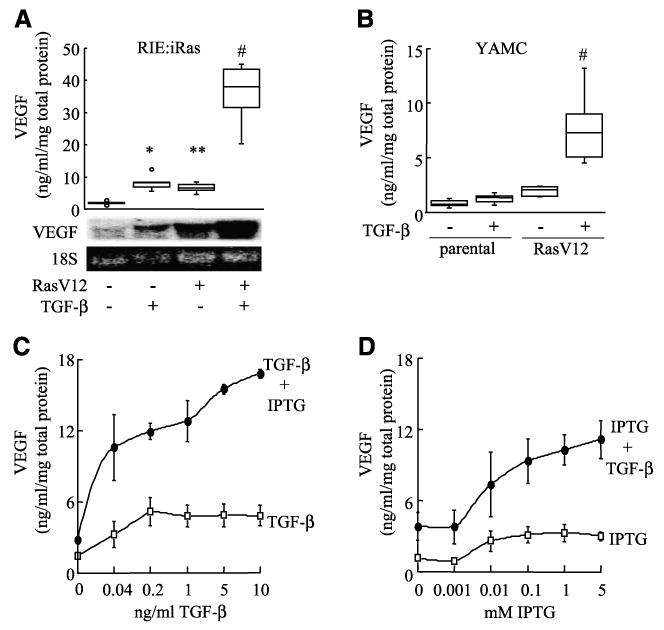
Oncogenic Ras and TGF-β synergistically increase VEGF expression in RIE:iRas and YAMC cells. A. RIE:iRas cells were treated with either vehicle, 5 mmol/L IPTG, 3 ng/mL TGF-β, or both IPTG and TGF-β for 24 h. VEGF protein levels were measured in conditioned medium by ELISA and normalized to total protein concentrations. Box plot shows data summarized from seven independently replicated experiments. *, P < 0.004, compared with untreated; **, P < 0.03, compared with untreated; #, P < 0.002, compared with all treatments. VEGF mRNA levels in RIE:iRas cells were visualized by Northern blot using a mouse VEGF165 cDNA probe and 18S rRNA visualized with ethidium bromide. Northern blot is representative of six separate experiments. B. YAMC and YAMC-Ras cells were treated with or without 5 ng/mL TGF-β for 24 h and then VEGF levels were measured in conditioned medium by ELISA and normalized to total protein concentrations. Box plot shows data from eight independently replicated experiments. #, P < 0.0001, compared with all samples. C and D. RIE:iRas cells were treated with IPTG and/or TGF-β at varying doses for 24 h and VEGF levels were measured in conditioned medium by ELISA and normalized to total protein concentrations. Cells were treated with or without 5 mmol/L IPTG and 0, 0.04, 0.2, 1, 5, or 10 ng/mL of TGF-β or cells were treated with or without 3 ng/mL TGF-β and 0, 0.001, 0.01, 0.1, 1, or 5 mmol/L of IPTG. Points, average of three independent experiments; bars, SE. Dose dependence and synergistic interaction were confirmed via multiple linear regression (all P < 0.0001).