Fig. 2.
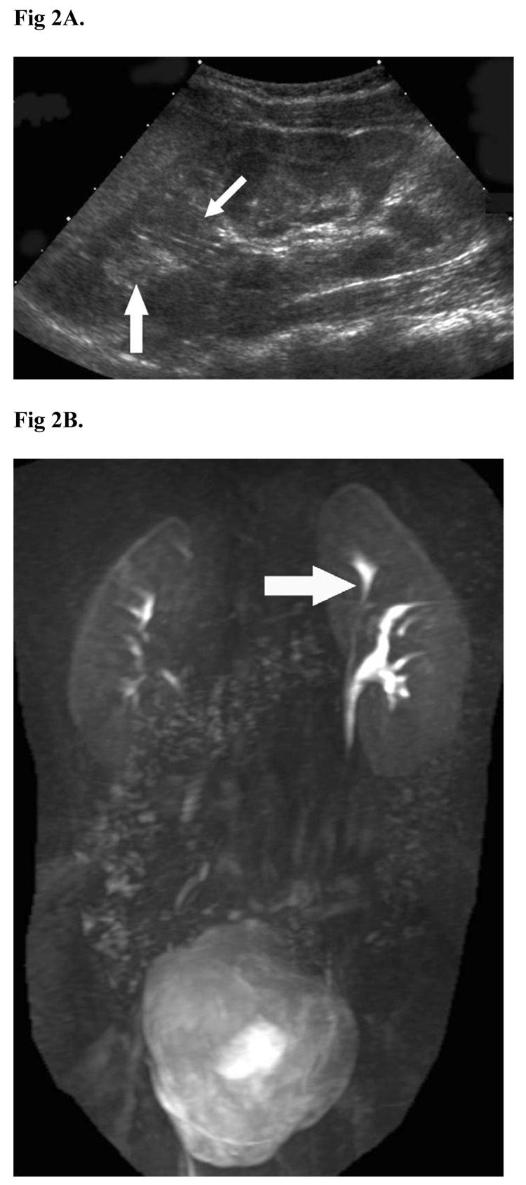
Five-year-old girl with perineal wetting. (A) Ultrasound shows an enlarged left kidney. Subtle duplication of the renal collecting system with intervening renal parenchyma (small arrow) dividing the renal sinus fat (large arrow) into two compartments was not initially diagnosed. (B) MR urogram shows a duplicated upper pole ureter (arrow).

