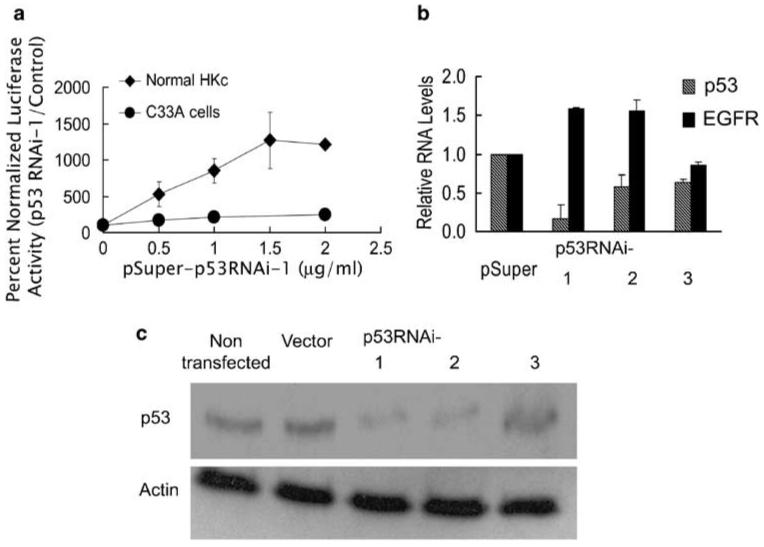
Figure 1.

Expression of a p53 RNAi results in induction of EGFR promoter activity in normal HKc. (a) EGFR promoter activity in normal HKc (◆) and C33A (●) in the presence of p53 RNAi. Normal HKc and C33A were transiently transfected with the pGL3-EGFR promoter construct (0.5 μg per well), pRL-TK (50 ng per well) and increasing concentrations of p53RNAi-1 or control pSuper plasmid. The total amount of DNA in the transfection mix was brought to 2.5 μg per well by addition of pSuper plasmid. Cells were harvested 48 h post-transfection for dual-luciferase assays. Data represent the averages±s.d. of four separate determinations using different normal HKc strains. (b) p53 and EGFR mRNA levels in normal HKc expressing p53 RNAi. Normal HKc were transfected with 5 μg per 60-mm dish of p53RNAi-1, p53RNAi-2, p53RNAi-3 or pSuper control plasmid and 1 μg per dish of pLXSN plasmid. Cells were selected with G418 (100 μgml-1) for a week, RNA was extracted and real-time RT-PCR was performed. The p53 and EGFR mRNA levels in the p53RNAi-transfected cells were normalized to RPLPO and expressed as a fold change compared with control (pSuper). Data represent averages±s.d. of triplicate determinations. (c) p53 protein levels in normal HKc expressing p53 RNAi. p53 or actin protein levels were detected by western blot analysis in extracts from normal HKc transfected with the various p53 RNAi constructs as described in (b). EGFR, epidermal growth factor receptor; HKc, human keratinocytes; RNAi, RNA interference; RPLPO, ribosomal protein large protein O; RT-PCR, reverse transcription-PCR.
