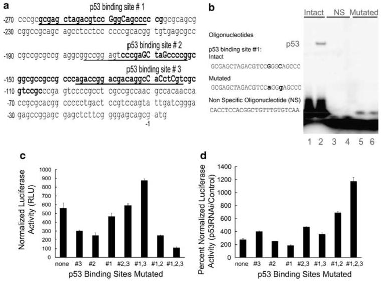
Figure 2.

Site directed mutations in p53 binding sites do not abolish induction of EGFR promoter activity by p53 RNAi. (a) Partial EGFR promoter sequence (nucleotides -270 to +3) containing the three regions where p53 binding sites have been previously identified, shown here in bold. The sequences we used in EMSAs are underlined. The specific nucleotides we mutated are shown in uppercase. (b) EMSAs were performed on synthetic oligonucleotides containing the p53 binding site 1, intact or mutated (M), and a non-specific control oligonucleotide (NS). These oligonucleotides were labeled with [γ- 32P] ATP and were incubated with nuclear extracts from C33A (lanes 1, 3 and 5) or UV-treated normal HKc (lanes 2, 4 and 6) and EMSA were performed as described in Materials and methods. (c) Basal activity and (d) p53RNAi-induced activity of the EGFR promoter p53 binding site mutants (single, double and triple) in transient transfections of normal HKc. Normal HKc were co-transfected with the indicated wild-type or mutated pGL3-EGFR promoter constructs (1.5 μg per well), along with pRL-Null (10 ng per well) (c) or with p53RNAi-1 or control pSuper plasmid (1 μg per well) and pRL-Null (10 ng per well) (d). Cells were harvested 48 h post-transfection for dual-luciferase assays. EGFR, epidermal growth factor receptor; EMSA, electrophoretic mobility shift assays; HKc, human keratinocytes; RNAi, RNA interference.
