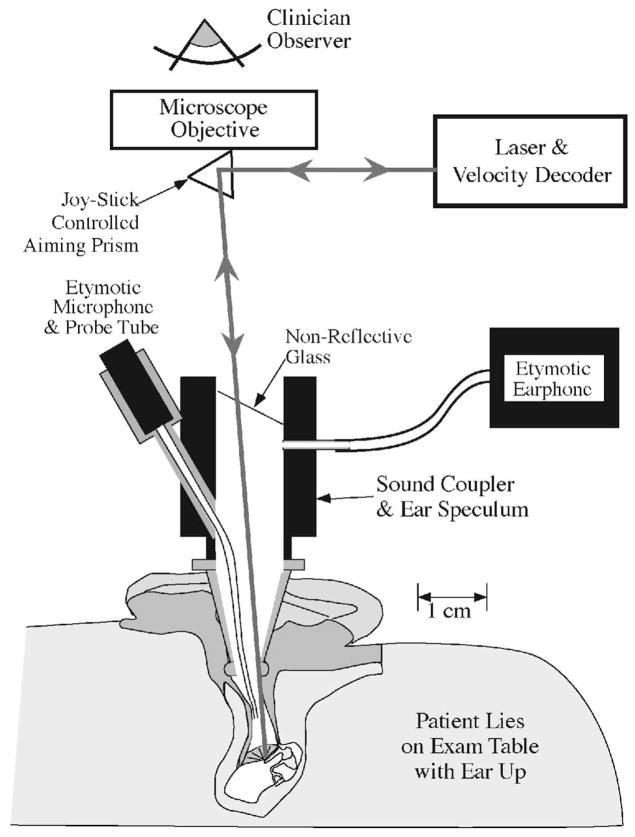
Fig. 1.
Laser Doppler vibrometry methods (after Whittemore, Merchant, Poon, & Rosowski, 2004). The patient lies supine on an examination table with the ear turned up. A speculum and operating microscope are used to examine the ear and observe the umbo of the malleus. A glass-backed sound coupler with attached microphone and earphone is attached to the speculum and the beam of the laser is focused through the glass onto the TM with the aid of a joystick-controlled prism. The light reflected from the TM travels back to the laser’s velocity decoder via the same optical path. A comparison of the frequencies of the transmitted and reflected light allows computation of the velocity of oscillation of the sound-driven TM.

