
Figure 2. Electron micrographs illustrating the diversity of synapses made by newly generated neurons.
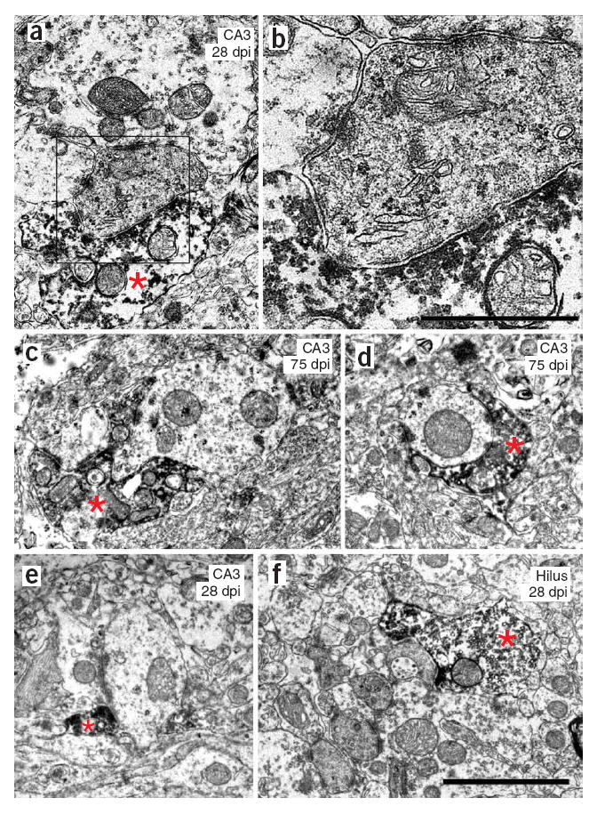
(a,b) GFP-positive mossy terminal synapsing on a thorny excrescence in the CA3 area at 28 dpi The boxed area is enlarged in b. (c) GFP-positive mossy terminal synapsing on a thin, spiny dendrite in the CA3 at 75 dpi. (d) Bouton en passant synapsing on a thin, aspiny dendrite in the CA3 area at 75 dpi. (e) Bouton en passant synapsing on a thin, aspiny dendrite in the CA3 at 28 dpi. (f) Mossy terminal synapsing on a thorny excrescence in the hilus at 28 dpi. Scale bars represent 2 μm in a and c–f, and 0.8 μm in b. Asterisks indicate GFP-positive axon terminal.