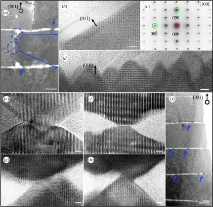
Figure 1.
Cross section TEM images demonstrate growth front of aragonite platelets in both vertical [001] and horizontal [010] directions. The nano-structures on the vertical (010) faces of platelets, as marked in (a), are magnified in (b) and (d) where (c) is the corresponding selected-area diffraction pattern showing the crystal orientation. (e–h) demonstrate that [001] outgrowths of nano-asperities from the top and bottom platelets are not exactly connected or epitaxial, even though they share the same crystal orientation as indicated by atomic lattice alignments. The atomic image in (d) reveals that those nano-growths are approximately 10–15 nm long and exhibit a larger aspect ratio than the growth structures found on the (001) faces of platelets. Scale bar, 3 nm in (b,d–h) and 200 nm in (a,i).

